Contents
What are Nudibranchs?
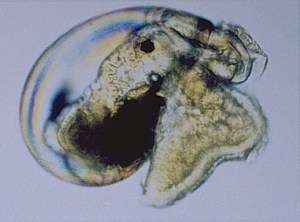
Nudibranchs are marine molluscs of the Class Gastropoda. Nudibranchs are commonly called as Sea slugs. An important character of nudibranchs is the absence of a calcareous shell in the adult forms. But their larval forms and many primitive forms still have shells.1 Nudibranch larvae are divided into three types based on the shape of larval shells.
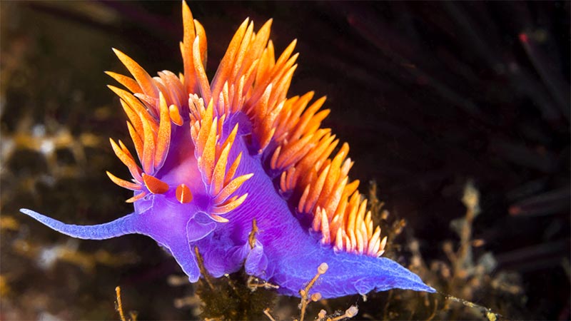
Striking feature of nudibranchs is their extraordinary colours and diversity of forms. They include one of the most colourful creatures on earth. Some species of nudibranchs exhibit aposematism through brilliant colouration.2 While some exhibit extreme camouflage.
View a video of a Nudibranch predating over other!
How did they get the name?
Nudibranchs or Nudibranchiae come from the Latin word nudus which means ‘naked’ and a Greek word brankhia meaning ‘gills’. Literally nudibranch means naked gills. The name is attributed due to the presence of anal or cloacal gills present in many forms (droids). Others (aeolids) have the presence of structures called as cerata which are similar to gills in appearance.
How are they classified?
Nudibranchs come under the Phylum Mollusca, Class Gastropoda and unranked clades Heterobranchia, Euthyneura and Nudipleura. Earlier they came under the subclass Ophisthobranchia. Now ophisthobranchia is not a valid clade due to its paraphyletic nature.
Nudibranchs are divided into two main groups, dorid nudibranchs and aeolid (spelled eolid) nudibranchs.
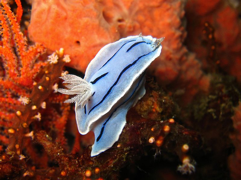
Dorids

In dorids, mantle is thick and extends over foot. Surface of the mantle usually bears tubercles that varies in size, shape and number. Dorids have branchial plume, a cluster of external gills on the posterior part of the body, near anus. In true dorids (ie. Dorididae, Rostangidae, Chromodorididae, etc. ) the gills can be retracted into a gill-pocket. These dorids are known as cryptobranch dorids as opposed to phanerobranch dorids in which the gills are contractile but not retractile into a pocket. Goniodorids. In some dorid families, the mantle is progressively reduced to a ridge around the side of the body, from which pallial tentacles or processes arise. These processes usually have coloured tips and contain defensive glands and have been shown to produce chemicals distateful to fish. These chemicals are often manufactured from similar chemical compounds in the bryozoan or ascidian prey, or may be the same molecules selectively re-secreted by the nudibranch.3
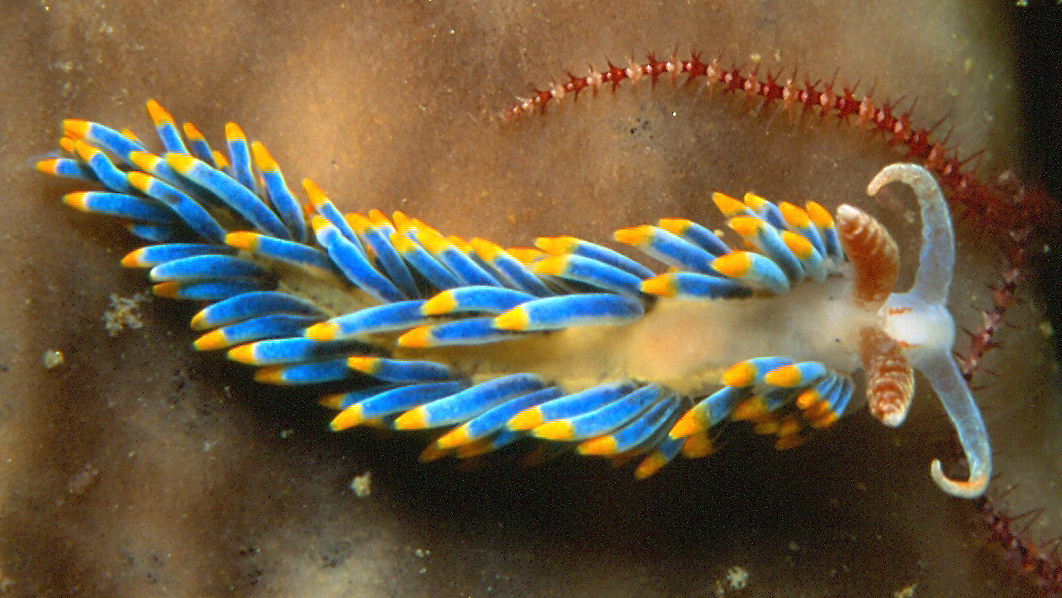
Aeolids
In aeolids, mantle is extended into long finger-like projections called cerata (singular: ceras). Cerata contain branches of digestive gland and often this is visible through the ceratal epidermis. In aeolids the tips of the cerata contain cnidosacs which usually store nematocysts (stinging cells) that are obtained from ingested cnidarian prey, such as hydroids, sea-anemones and soft corals. If disturbed, the nudibranch is capable of discharging these stinging cells through a terminal pore in the ceras; this is an effective deterrent to predatory fish.3
What are the anatomical features of nudibranchs?
Symmetry: They exhibit bilateral symmetry externally. But the placement of many internal organs does not exhibit any symmetry. The presence of genital openings on the right side of the body shows their assymetrical origin. This is because, they have undergone detorsion. In gastropods, torsion is the rotation of visceral mass with respect to head and foot. Ancestors of nudibranchs exhibited torsion. They have secondarily lost this character and is called as detorsion.
Shell
Most of the nudibranchs do not have a calcareous shell around the body. The loss of the shell gave them the opportunity to experiment with all sorts of body shapes and to experiment with the brilliant and spectacular colour patterns.1
Mantle Cavity and Gills
Another important character is the absence of a mantle cavity. Mantle cavity acts as a respiratory chamber in molluscs. A reduction in respiratory chamber is compensated with the formation of external gills and cerata (which function as secondary gills) in these organisms.
Even though gaseous exchange occurs through the general body surface, gills are the most important respiratory organ in nudibranchs. In dorids, gills consist of several feather-like structures that encircle the anus. This structure is termed the branchial plume and is situated in the dorsal region in posterior part of the animal. In some droids, these gills can be retracted into gill pockets. In aeolids, cerata function as gills. The ceratal epidermis is thin enough to enable gaseous exchange with surrounding water.
Eye
The head region of nudibranchs has a photosensitive organ above the brain, namely ocelli. Even though they cannot form an image, they help the slug distinguish dark and light.4
Rhinopores
Head also bears a pair of sensory tentacles called rhinophores. These are chemosensory (smell, taste) in function. In many dorid nudibranchs the rhinophores can be retracted into a basal sheath. The shape of the rhinophores varies greatly from one species to another.
Oral tentacles
Many nudibranchs also have a pair of oral tentacles on either side of the mouth. They are probably chemoreceptors or touch receptors.3
Defence Systems
In molluscs, shell acts as the primary defence system. But shells can be disadvantageous for movement and growth. Loss of shells gave nudibranchs the freedom to evolve various body shapes and comparatively fast movement. These advantages compelled them to evolve a series of mechanisms to protect themselves from their predators. These defence systems, real chemical and biological weapons, could have been either a post-adaptation to shell loss, either a pre-adaptation that made the shell loss possible.5
Acid glands and spicules
In many dorids acid glands and/or spicules are incorporated in the mantle tissue, which are mainly defensive in function. In many dorid families, the mantle is progressively reduced to a ridge around the side of the body, from which pallial tentacles or processes arise. These processes usually have coloured tips and contain defensive glands and have been shown to produce chemicals distateful to fish. These chemicals are often manufactured from similar chemical compounds in the bryozoan or ascidian prey, or may be the same molecules selectively re-secreted by the nudibranch.
Autotomy
There are a number of species that have evolved the ability to drop cerata from body, which can be rapidly regenerated, to confuse predators and escape. This is called autotomy.
Camouflage
In many nudibranchs, both the texture and the colour of the match the colour and the texture of the prey or the substrate they live on. It is called cryptic coloration. Nudibranchs obtain the pigments from their diets or sometimes they produce pigments themselves. In many cases, slugs can vary their colour as they change the food source
Mucus Coating
Many nudibranchs secrete musus which is resistant to nematocysts. They are supposed to contain some inhibitory agents.
Aposematic colouration
Some nudibranchs have a bright and spectacular colour patterns. These colours are used to warning to potential predators. This is known as aposematic colouration. Normally these slugs contain distasteful or even toxic compounds. Nudibranchs obtain the pigments from their diets, sponge or cnidarians, or even they can produce de novo themselves.
Cnidosacs
Many species of aeolid nudibranchs collect and store undischarged nematocysts, for use in their own defense. The nematocysts are stored in special sacs or cnidosacs located in the tips of the cerata. Each cnidosac is lined with a single-layered epithelium composed mainly of phagocytosing cells called cnidophages. Nematocysts that are eaten are sorted in the stomach and shunted into the diverticlua of the digestive gland that extend into the cerata. From there they pass through a narrow canal that opens into each cnidosac. On entry into the lumen of the cnidosac the nematocysts are phagocytosed by the cnidophages. Within the cnidophages the nematocysts may lie parallel with one another, or be arranged in circles or bouquets depending upon the species.
When nematocyst-bearing aeolids are disturbed or irritated, they curl their bodies and bristle their cerata. Special muscles surrounding the cnidosacs squeeze the nematocysts from their sacs and enclosing cnidophage cells. They are forced out of cnidopore at the tip of the ceras. Some of these nematocysts penetrate the predator and it withdraws.6
References
-
1.W.B. R. Larval shell. The Sea Slug Forum. http://www.seaslugforum.net/showall/larvshel. Accessed November 28, 2016.
-
2.Ritson-Williams R, Paul V. Marine benthic invertebrates use multimodal cues for defense against reef fish. Marine Ecology Progress Series. 2007;340:29-39. doi:10.3354/meps340029
-
3.Nudibranchs : Anatomy . Nudibranchs of the British Isles. http://www.seaslug.org.uk/nudibranchs/anatomy.html. Accessed November 28, 2016.
-
4.W.B. R. The nudibranch head. The Sea Slug Forum. http://www.seaslugforum.net/find/headeudo. Accessed November 28, 2016.
-
5.Feature Article: Nudibranchs. Beautiful but Dangerous Marine Creatures. http://www.advancedaquarist.com/2007/11/aafeature2#section-1. Accessed November 28, 2016.
-
6.Kälker H, Schmekel L. Structure and function of cnidosac of Aeolidoidea (Gastropoda, Nudibranchia). Zoomorphologie. 1976;(86):41-60.


Leave a comment