General Zoology
Common pet diseases and how to prevent them
We love our pets so much that if their health starts to decline, it can be devastating. Learn what you can do to...
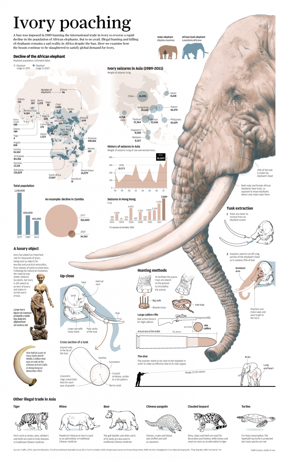
Everything you need to know about ivory poaching
A ban was imposed in 1989 banning the international trade in ivory to reverse a rapid decline in the population of African elephants....
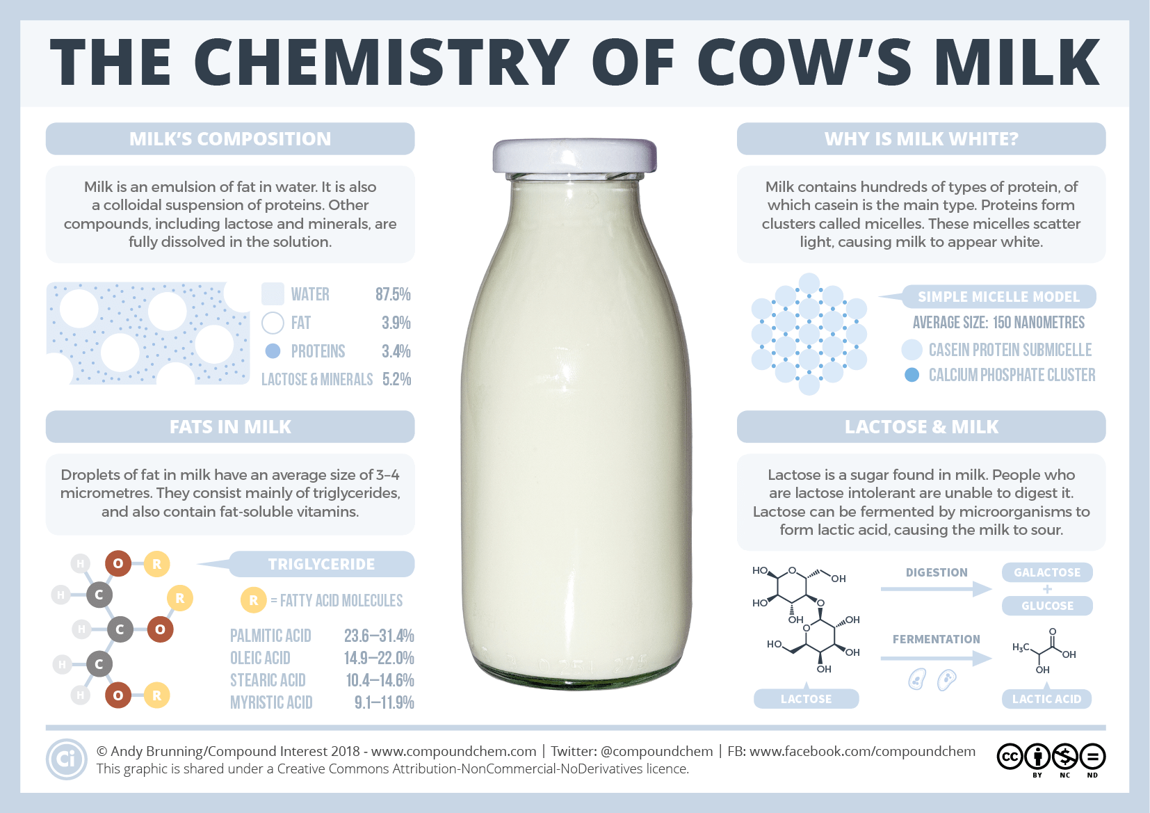
The chemistry of milk
In milk, proteins cluster together to form structures called micelles. They grow from small clusters of calcium phosphate, which held the proteins together....
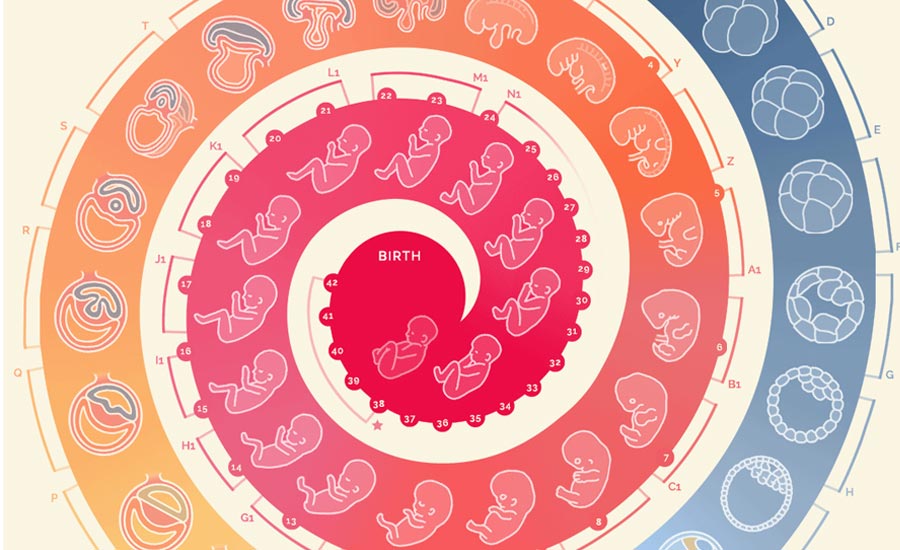
How to build a human
Infographic showing human development from fertilization to birth.

Top 20 Zoology blogs for students and educators
Useful selection of top zoology blogs for students and teachers of zoology. These handpicked blogs range from General zoology, Evolution, Marine biology, cryptozoology...
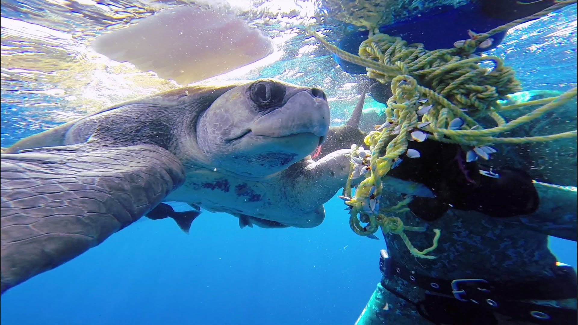
Sea turtle thanks divers for rescuing
Colin Sutton and Cameron Dietrich were diving off the coast of Mexico when they found a sea turtle tangled up in the...
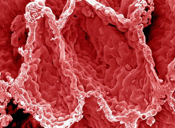
These electron microscopic images of human body will astonish you
Artery Coloured scanning electron micrograph (SEM) of a a small artery (blood vessel) cut open with RBCs (red coloured) rushing outside. Blood Clot...